Glaucoma.
Glaucoma is one of the leading cause of blindness throughout the world,
affecting approximately 2% of the population over the age of 40. Unlike
many other eye diseases, conditions or problems, glaucoma
begins without
any symptoms or obvious loss of vision. Often called the sneak thief
of sight, glaucoma can destroy sight without causing any pain or obvious
symptoms until it is too late. Thus, early detection of glaucoma
is extremely important. This disease can often be successfully treated,
and blindness prevented, when diagnosed early enough. We invite you to make an appointment and learn more: 480-981-6111.
What is Glaucoma?
A complex eye disease, glaucoma is not simply an elevated intraocular
pressure. Glaucoma is actually a broad term that is
used to characterize a range of eye conditions that damage the optic
nerve and potentially cause loss of vision. Glaucoma
therefore is a disease of the optic nerve, the vital nerve bundle that
carries images to your brain so you can see.
Glaucoma usually affects both
eyes, but can progress more rapidly in one eye than in the other. Involvement
of just one eye primarily occurs when glaucoma is brought on by factors
such as a prior injury or the use of steroids in that eye.
How Glaucoma Works
The eyeball is basically a rigid sphere filled with aqueous humor.
In the normal eye, there is a constant production and drainage of
this fluid. This production and drainage is balanced so as to maintain
a “normal' intraocular
pressure (IOP). As
the total amount of fluid within the eye increases, so does the pressure.
Many people relate glaucoma to increased pressure inside the eye,
although that is not the only cause, but is a common cause of glaucoma.
The higher the pressure inside the eye, the greater the chance of
damage to the optic nerve.
Imagine that a healthy eye is like a water
balloon attached to a faucet that is permanently dripping water.
The balloon has a drainpipe that slowly filters the water out,
at a constant rate, allowing the balloon to stay perfectly full without
overextending it. However, if the drainpipe were clogged, the water
would collect inside the balloon and over-inflate it, creating too
much pressure. Eventually, the most fragile part of the balloon would
become damaged and give way.
In your eye, the liquid is called aqueous humor and constantly
flows in and out, not as a part of the tears on the outer surface,
but rather coming from inside your eye and leaving through a drainpipe
called the angle (because of its angular shape). If the angle
becomes blocked, the volume of fluid inside the eye goes well beyond
normal levels, putting too much pressure on the fragile
optic nerve
and damaging it (see illustration at the top of this page).
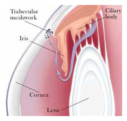
The diagram at the right is of the front part of the eye, in cross section,
to show the filtering, or drainage angle. The angle is between the cornea
and the iris, which join each other right where the drainpipe (called the trabecular
meshwork)
is located. The purple arrow in the diagram shows the flow of the
aqueous fluid from the ciliary body, through the pupil, and into
the drainage channels.
Glaucoma can affect anyone. While high pressure
in the eye is considered
the primary reason for glaucoma, even patients with normal
eye pressure have been known to contract this disease. At East
Valley Ophthalmology, your eye doctor puts together many kinds of
information to determine your risk for developing glaucoma. If
detected early, loss of vision can most often be prevented.
Risk
Factors of Glaucoma
Factors that
increase a person's risk generally include:
· Increased Intraocular
Pressure (IOP) - Anyone who has been found to have an elevated
intraocular pressure at a glaucoma screening or as part of
a general eye examination is considered to be at risk for developing
glaucoma.
· Increasing Age -
The incidence of glaucoma increases as we get older. Typically
the incidence of glaucoma becomes much more noticeable above the
age of 40 years old. This is why routine eye
examinations with
glaucoma evaluation are recommended every 2 years above the age
of 40, if there is no other family or medical history, and more
often if there is a preexisting history of glaucoma in the family
or other predisposing heath factors.
· Race -
African-Americans to have certain genetic factors that cause
a higher likelihood of developing glaucoma.
· High Blood
Pressure - Those patients
who are being medically treated for high blood pressure may be
at greater risk for glaucoma due to the lowering of the blood
pressure within the optic nerve.
· Family History
of Glaucoma - Family history
of glaucoma is a very significant risk factor. If any other family members
have been diagnosed with glaucoma, your risk of developing glaucoma increases
considerably. This is particularly true for siblings of glaucoma patients,
who have
a 5-fold increase in risk for developing glaucoma.
· Diabetes - Anyone being treated
for diabetes is considered to be at greater risk for glaucoma due to the general
circulation problems associated with diabetes.
· Myopia (nearsightedness) - In general patients
who are nearsighted have anatomical features that may increase
the risk of glaucoma.
· Long-term Steroid
Treatment
· Injury/Trauma
To The Eye
Your eye doctor will weight all of these factors before deciding whether
you need treatment for glaucoma, or whether you should be monitored
closely as a glaucoma suspect, meaning your risk of developing glaucoma
is higher than normal, and you need to have regular eye
examinations
to detect the early signs of damage to the optic nerve.
Types
of Glaucoma
The eye's drainage system,
which lies in-between the cornea and
the iris, is called the angle,
named for its angular shape. Therefore, you see the word angle in
the different glaucoma names. While there are many types
of glaucoma, they primarily fall into two groups, differentiated
according to whether the angle is open (able to accept fluid) or
closed (fluid is blocked from entering).
Open Angle Glaucoma
Open angle glaucoma is chronic —
develops over time.
Closed Angle Glaucoma (Angle-Closure Glaucoma)
Closed angle glaucoma is acute — can occur suddenly.
Primary Open Angle Glaucoma PAG
By far, the most frequently diagnosed type of Glaucoma
in the United States is primary open angle glaucoma (POAG). Patients
with primary open angle glaucoma typically demonstrate an increase
in intraocular pressure (IOP) upon routine measurement. The increased
intraocular pressure results from either too much aqueous humor
being produced or too little being drained. This
fluid buildup within the closed space of the inside of the eye elevates
the pressure. It is this raised pressure that can cause permanent
changes and even damage to the optic nerve resulting in vision loss.
Since the optic nerve is the connection between the retina and the
brain and is responsible for communicating visual images, once the
optic nerve is damaged, vision can no longer function. This is why
it is so important to monitor, detect and control intraocular pressure.
If left untreated, an elevated intraocular pressure may, over
time, cause slow progressive, permanent damage to the optic nerve
that can result in blindness.
Initially, open-angle glaucoma has no symptoms. There
is no pain or noticeable change in vision. Loss of peripheral (vision
off to the side) is the earliest symptom. Left untreated the field
of vision will continue to narrow, leading to tunnel vision, eventually
to blindness.
Closed Angle Glaucoma (Angle-Closure Glaucoma)
Acute angle-closure
glaucoma is an emergency and should be treated immediately.
Closed-angle glaucoma occurs when fluid cannot escape because the
drainage angle is closed off, which causes pressure to build up suddenly.
This often causes noticeable pain.
Angle
closure glaucoma is found much less frequently than open angle glaucoma,
but it has the ability to produce considerable vision loss in a short
period of time. While there can be a number of causes of closed angle
glaucoma, it is most often caused by anatomical changes within the
internal structures of the eye.
Acute closed angle glaucoma is structurally different
from open angle. With closed angle glaucoma, the fluid in the eye
cannot get to the drainage meshwork (trabecular meshwork) because
the clogging is before the meshwork. In contrast, with open angle
glaucoma the clogging is within the drainage meshwork itself.
The
trabecular meshwork is actually a tiny tissue filter that, if blocked
by a change in size or shape of the tissue, will cause the intraocular
pressure to elevate. In instances where the meshwork becomes blocked
abruptly, it will cause a sudden rise in the intraocular
pressure. This sudden rise in pressure can cause pain, redness, blurred
vision and if left untreated permanent loss of vision.
Closed angle glaucoma is considerably more common in farsighted
eyes, which tend to be smaller, and in patients between the ages
of 45-60 years of age where the crystalline lens is beginning to
swell.
During your general eye exam, if your eye doctor
observes or measures a narrowed angle, he will perform an
additional examination procedure called Gonioscopy to fully visualize
the meshwork and the angle in order to carefully assess your predisposition
to angle closure glaucoma. This test is performed by placing a special
contact lens on the eye and then using the slit lamp biomicroscope
to fully examine the meshwork and the angle. In the event that you
are at risk for angle closure glaucoma, or in the event that you
have acute
angle closure glaucoma, the most effective form of treatment is to
use a laser to produce a small opening or hole in the iris so that
aqueous humor can quickly and efficiently drain from the eye by preventing
the trabecular meshwork from being blocked.
Secondary Glaucoma
Glaucoma resulting from congenital, ocular or systemic conditions
represent secondary glaucoma.
The six most common forms of secondary glaucoma:
-
Exfoliation syndrome
-
Pigmentary glaucoma
-
Neovascular glaucoma
-
Lens induced glaucoma
-
Glaucoma accompanied by ocular inflammation
-
Trauma induced glaucoma
Diagnosing Glaucoma
Routine eye examinations are
mandatory because, in its early stages, glaucoma usually has no symptoms.
In order to be controlled, glaucoma must be diagnosed early. It is
a lifelong disease, so those afflicted must be compliant for the
rest of their lives and religiously keep to their scheduled eye exams
and with their medication regimens.
A glaucoma exam includes:
·
Measuring your intraocular pressure (tonometry)
·
Inspecting the drainage angle of your eye (gonioscopy)
· Evaluating any
optic nerve damage (ophthalmoscopy)
· Testing the
visual field of each eye (perimetry)
Some of these tests may not be necessary for every person. You may need to repeat these tests on a regular basis, to determine if glaucoma damage is increasing over time.
When should you have a glaucoma exam?
· Under age 45
With risk factors = every 2 years.
Without risk factors = every 4 years.
· Over age 45
With risk factors = every year.
Without risk factors = every 2 years.

TOP
Treatment Of Glaucoma
As a rule, damage caused by glaucoma cannot be reversed. Therefore, the goal
in the management of glaucoma is to reduce the intraocular pressure to the
point whereby the remaining healthy nerve fibers are able to receive proper
nourishment and maintain the remaining function. Proper treatment can keep
the intraocular pressure within normal range and therefore prevent or retard
further nerve damage and visual loss.
Eye Drops for Glaucoma
Open angle glaucoma is usually controlled with eye drops taken several times
a day , sometimes
in combination with pills. There are many types of eye drops available that
can lower the intraocular pressure. By using a single type of medication
or sometimes 2 eye drops in combination, more than 80% of the patients
with open angle glaucoma can be successfully treated.
One or more
types of eye drops may have to be taken up to several times a day
in order to be effective. These medications are used to prevent damage
to the optic nerve by decrease eye pressure, either by slowing the
production of aqueous fluid within the eye or by improving the flow
leaving the eye. Recently there have been a few brand new medications
which show great promise for more effectively than others. In order for these
medications to work, you must take them regularly and continuously as they
were prescribed. The vital key to the success of medication therapy
is patient compliance.
Laser Surgery for Glaucoma
Some patients experience side effects of these eye drops and it makes the
use of eye drops a poor treatment option. Also, some patients are unable to
achieve adequate control with eye drops alone and require laser treatment in
addition to the eye drops in order to maintain control. The laser is
usually used in one of two ways"
Open Angle Glaucoma
Treatment for open angle glaucoma involves the
drain itself. In open-angle glaucoma, the laser is
used to enlarge the drain (argon laser trabeculoplasty) to help control eye
pressure.
Closed Angle Glaucoma (Angle-Closure Glaucoma)
In angle-closure glaucoma, the laser
creates a hole in the iris (iridotomy) to improve the flow of aqueous fluid
to the drain.
Each type of treatment has its benefits and potential complications. At East
Valley Ophthalmology, our physicians will go over them with you in detail and
answer all your questions and concerns.
Surgical Treatment of Glaucoma
For a small number of patients,
it is still not possible to achieve good stable control and stop the
progression of glaucoma. For these patients there are surgical procedures
including removing a tiny piece of the trabecular meshwork or even implanting
a microscopic glaucoma valve that can be performed to help reduce and
stabilize the intraocular pressure and prevent vision loss.
When operative surgery is needed to control glaucoma, your eye surgeon
creates a new drainage channel (bleb) through which the aqueous fluid
can leave the eye, thereby lowering the IOP.
Fortunately, technology has improved significantly for both the medication
and laser treatment alternatives in glaucoma, so that only a very few
individuals need ever progress to the point of needing surgery. The
key to a lot of these successes however, lie in prevention; specifically,
the earlier that glaucoma can be diagnosed, the more effective the treatment
through either medications or laser.
The eye specialists of East Valley Ophthalmology perform advanced
technology diagnostic testing and treatment, as well as taking
the time necessary to provide each patient with information needed
to fully understand their condition and to achieve their best possible
visual outcome.
If you would like further information, please call our office at:
480-981-6111
East Valley Ophthalmology
Eye Doctors - Mesa, ArizonaIf you or a family member
or friend have not had a recent routine eye examination, have a specific eye condition that needs addressing, or are looking for
an eye specialist or professional eye consultant please take a moment to Request an Appointment.
|