Pterygium Removal
No-Stitch Pterygium Surgery —
More Comfortable Removal — Shorter Recovery!*
We invite you to make an appointment and learn more: 480-981-6111.
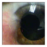
Pterygium (pronounced "tur-RIDGE-ium")
is a pinkish-yellow, triangular-shaped benign tissue growth starting
from the nasal area of your eye and grows toward the cornea. As
a pterygium grows, it can be varied in its appearance from small
and pink to large and angry red with symptoms of dry eye. Eventually,
it may cause visual disturbances by disrupting the normally smooth
surface of the cornea. In severe cases, a pterygium can block a
patient's vision altogether.
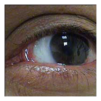
Before Pterygium Removal  After
Pterygium Removal Dr. Jonathan Kao performs state-of-the-art pterygium removal
surgery and is well known for his "no-stitch" surgical
technique and instrumentation. He performs a complete excision
of this lesion using no stitches during this surgery, and instead
applies a special surgical glue*. This highly specialized method
for pterygium removal has raised the bar in looks and comfort for
patients with pterygium. Patients from all over the country come
to East Valley Ophthalmology for the expertise of Dr. Kao and his
method to remove pterygiums.
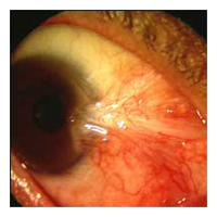
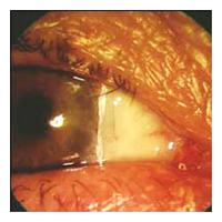
Before Pterygium Removal After
Pterygium Removal
Causes of Pterygium
Pterygia are more common in sunny climates and in the 20-40 age group. Scientists
do not know what causes pterygia to develop. However, since people who have pterygia
usually have spent a significant time outdoors, many doctors believe ultraviolet
(UV) light from the sun may be a factor. In areas where sunlight is strong, wearing
protective eyeglasses, sunglasses, and/or hats with brims are suggested. While
some studies report a higher prevalence of pterygia in men than in women, this
may reflect different rates of exposure to UV light.
Preventing Pterygium
The best method of preventing pterygium is to regularly wear UV
400 rated sunglasses when outdoors in sunny conditions. Sunglasses
with a wrap-around design provide better protection than those with
large gaps between the sunglass frame and the skin around the eyes.
Wearing a hat with a wide brim provides valuable additional protection.
Treating Pterygium
In mild cases, pterygium redness and discomfort can be controlled
with lubricant eye drops (artificial tears). When symptoms of redness,
irritation, or blurred vision are resistant to conservative treatment,
or when vision is affected by progressive growth of a pterygium,
surgery is considered.
Pterygium Surgery
History of Pterygium Surgery
In pterygium surgery, the abnormal tissue is removed from the cornea
and sclera (white of the eye). Over the years, pterygium surgery has
evolved significantly, and modern pterygium surgery has a
significantly higher success rate than conventional surgery.
In traditional "bare sclera" pterygium removal, the underlying
white of the eye (sclera) is left exposed. Healing occurs over two
to four weeks with mild to moderate discomfort. Unfortunately, the
pterygium may grow back in up to 50% of patients. In many cases,
the pterygium grows back larger than its original size.
Over the years, surgeons have used several different techniques
to lessen the likelihood of pterygium recurrence, including radiation
treatment and the use of "antimetabolite" chemicals that prevent
growth of tissue. Each of these techniques has risks that potentially
threaten the health of the eye after surgery, including persistent
epithelial defects (ulceration in the surface of the eye), and corneal
melting.
Conjunctival Graft with Stitches
Most cornea specialists today perform pterygium surgery and cover the
exposed area with a graft to reduce the risk of recurrence. There are
two main sources of this graft. One is your own body; in this
technique a thin layer of the white membrane, or conjunctiva, covering
the eye is removed from beneath the upper eyelid. This is then moved
over to fill the area where the pterygium once occupied. Although the
procedure requires more surgical skill and time than traditional
surgery, this "auto-graft" (self-transplant) helps prevent re-growth
of the pterygium by filling the space where abnormal tissue would have
re-grown.
The autograft is held in place with tiny stitches that may dissolve
after a few weeks or can be removed in the surgeon's office. Stitches,
however, may cause discomfort and a foreign body sensation.
Amniotic Membrane Graft
The other source of covering is amniotic membrane, which is a unique
membrane derived from the submucosa of the placenta, the structure
that provides nutrition to a developing fetus. The tissue is donated
by consenting mothers and screened for infectious diseases according
to FDA standards. Because of its' origin, amniotic membrane is filled
with natural molecules that are anti-inflammatory, anti-scarring, and
promote wound healing.
Regardless of the covering used, the desire for a quicker, more
painless recovery has led to the development of no-stitch pterygium
surgery.
No-stitch Pterygium Surgery*
No-stitch pterygium surgery allows most patients to return to work
within one or two days of surgery. Research studies have shown that
patients undergoing no-stitch surgery had significantly less pain and
discomfort after surgery than those having traditional surgery. The
no-stitch technique also reduced surgery time by 20-30%.
Technique for No-stitch Pterygium Surgery
In no-stitch surgery, the patient is lightly sedated to ensure
comfort, and the eye is completely numbed, so there is no way to see
the surgery occurring and no sensation of discomfort. The abnormal
tissue is removed and replaced with a thin autograft or amniotic
membrane. Over the next 2-4 weeks, the eye gradually returns to a
normal appearance.
No-stitch surgery is made possible by the use of modern tissue
adhesive. Composed of clotting proteins normally found in human blood,
tissue adhesive allows the surgeon to secure a conjunctival autograft
in seconds rather than minutes. After about one week the tissue
adhesive dissolves with no residue, leaving the eye to heal
comfortably. Although tissue adhesive is derived from human blood
products, no cases of blood borne infection have ever been reported
among millions of patients treated with this material in heart and
lung surgery.
*Fibrin tissue adhesive is a drug approved by the FDA for abdominal surgery. Although its use in eye surgery has not yet been specifically approved by the FDA, our research suggests that this technique is a safe and effective alternative to eye surgery using stitches.
The eye specialists of East Valley Ophthalmology perform advanced
technology diagnostic testing and treatment, as well as taking
the time necessary to provide each patient with information needed
to fully understand their condition and to achieve their best possible
visual outcome.
If you would like further information, please call our office at:
480-981-6111
East Valley Ophthalmology
Eye Doctors - Mesa, ArizonaIf you or a family member
or friend have not had a recent routine eye examination, have a specific eye condition that needs addressing, or are looking for
an eye specialist or professional eye consultant please take a moment to Request an Appointment.
|