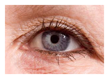
Eye Examinations.
Make an appointment for
your eye examination at East Valley Ophthalmology and read this section
to understand and prepare for your eye exam. Call now:
480-981-6111
Prepare for Your Eye Exam:
As a new patient, please download New
Patient Forms ahead of time and either bring them
with you or FAX them to our office prior to your appointment. Our FAX
number is: 480-985-2426 Before your appointment, find out if you will be given
drops to dilate your eyes. Although the effects of dilating drops are
temporary, you may need to arrange for someone else to drive or to
pick you up after your exam.
In addition, please be prepared to answer questions regarding your
health history, vision-related concerns and expectations. This will
help us understand the health of your eyes, your risk of eye disease
and vision problems, and allow us to make the best possible evaluations
and suggestions regarding your current situation. Ideally, make
a written list of this information ahead of time and bring it with
you.
Include:
- Your overall health history and that of your family, especially
relating to any eye diseases, such as cataracts or glaucoma.
Include any health problems you've had in recent years.
- Your family history of diabetes, high blood pressure, heart
disease or any other health problems that can affect the whole body.
- Any allergies you have to medications, food or other substances.
- Eye problems you have now, plus any you've experienced in the past.
- Bring along all medications
you are taking, even if they appear to have nothing to do with your
eyes. Some drugs have side effects that could affect your eyes.
- Make a list of questions you have about your condition and bring it with
you.
- Know your health insurance coverage for
vision care. Not all insurance pays for routine vision exams such as
for eyeglasses. Others have limitations on anything outside routine
procedures and, should you need any, you would then be personally responsible.
- If you wear glasses or contacts now and how satisfied you are with
them.
-
If you wear contact lenses or glasses, bring them to your appointment. We will
want to make sure your prescription is the best one for you. If you wear contact
lenses, be prepared to remove them. Tests that use orange dye (fluorescein)
to temporarily color your eye may permanently dye your contact lenses.
During the Eye Exam:
Part of the examination, such as taking your medical history and the
initial eye test, may be performed by a technician who assists your
doctor. This information will be reviewed by your eye
doctor prior to examining you.
A complete eye exam involves a series of tests designed to evaluate
your vision and check for eye diseases. All of it is painless. Your
doctor may use odd-looking instruments, aim bright lights directly
at your eyes and request that you look through a seemingly endless
array of lenses. Each test evaluates a different aspect of your vision.
We want you to feel completely comfortable with your eye exam.
If some part of the process is confusing to you, please ask for further explanation.
Should you have further questions later, don't hesitate to call us. There
are no foolish questions, only ones you want answered, and we encourage
you to ask. Plus, there are usually printed materials about your condition
that you can take home.
Common Tests During an Eye Exam:
Eye exams involve more than testing your vision and, if
you need glasses or contacts, determining how strong your correction
should be. Additional tests assess the appearance and function of
all parts of your eyes.
Eye muscle test
This test examines the muscles that control eye
movement, looking for weakness or poor control. Your eye doctor
looks at your eyes as you move them in six specific directions and
as you visually track a moving object, such as a pen.
Visual acuity test
This test measures how clearly you can see from
a distance. Your doctor will ask you to identify different letters
of the alphabet printed on a chart positioned usually 20 feet away.
The lines of type get smaller as you move down the chart. You cover
one eye and read aloud, then cover the other eye and read aloud.
Refraction assessment
"Refraction" refers to how light waves are
bent as they pass through your cornea and lens. A refraction assessment
helps your doctor determine a corrective lens prescription that
will give you the sharpest vision. If you don't need corrective
lenses, you won't have a refraction assessment.
Your doctor may use a computerized refractor to measure your eyes
and estimate the prescription you need to correct a refractive error.
Or he or she may use a technique called retinoscopy. In this procedure
the doctor shines a light into your eye and measures the refractive
error by evaluating the movement of the light reflected by your
retina.
Your eye doctor fine-tunes this refraction assessment by asking
you to look through a Phoroptor, a mask-like device that contains
wheels of different lenses, and judge which combination gives you
the sharpest vision. By repeating this step several times, your
doctor finds the lenses that give you the greatest possible acuity.
Visual field test (perimetry)
Your visual field is the area directly in
front of you that you can see without moving your eyes. The visual
field test determines whether you have difficulty seeing in any
areas of your peripheral vision — the areas on the side of
your visual field. There are a few different types of visual field
tests:
- Confrontation visual field exam. Your eye doctor sits directly
in front of you and asks you to cover one eye. You look directly
at your eye doctor while he or she moves his or her hand in and
out of your visual field. You tell your doctor when you can see
his or her hand.
- Tangent screen exam. You sit a short distance
from a screen and stare at a target at its center. You tell your
doctor when you can see an object move into your peripheral vision.
- Automated perimetry. Your eye doctor uses a computer program that
flashes small lights as you look into a special instrument. You
press a button when you see the lights.
By gathering information based on your responses to one or more
of these tests, your eye doctor makes a map of your peripheral vision.
If you aren't able to see in certain areas, your eye doctor uses
the map to help diagnose your eye condition.
Slit-lamp examination
A slit lamp is a microscope that enlarges
and illuminates the front of your eye with an intense line of light.
Your doctor uses this light to examine the cornea, iris, lens and
anterior chamber of your eye.
When examining your cornea, your doctor may use eye drops containing
fluorescein (flooh-RES-ene) dye. The orange dye spreads across your
eyes to help your eye doctor detect tiny cuts, scrapes, tears, foreign
objects or infections on your cornea. Your eyes' tears will wash
the dye away.
Retinal examination (ophthalmoscopy)
A retinal examination — sometimes
called ophthalmoscopy or fundoscopy — examines the back of
your eye, including your retina, optic disk and the underlying layer
of blood vessels that nourish the retina (choroid). Usually before
your doctor can see these structures, your pupils must be dilated
with special eye drops The eye drops may sting briefly and might
cause a medicinal taste in your mouth as the medication drains from
your tear ducts into your throat.
After administering eye drops, your eye doctor may use one or more
of these techniques to view the back of your eye:
- Direct examination. Your eye doctor shines a beam of light
through your pupil and uses an ophthalmoscope to see the back of your
eye. Sometimes eye drops aren't necessary to dilate your eyes before
this exam. You might see afterimages when your eye doctor stops shining
the light in your eyes. This is normal and will go away.
- Indirect
examination. For this exam you might lie down or recline in a chair.
Your eye doctor will hold each eye open and examine it with a bright
light mounted on his or her forehead — a bit like a miner's
lamp. This exam lets your eye doctor see your eye in great detail
and in three dimensions. Since this light is brighter than that
in a direct examination, you are more likely to see afterimages,
but they disappear quickly.
- Slit-lamp exam. In this exam your
doctor uses the slit lamp along with the ophthalmoscope to look
at the back of your eye. The slit lamp reveals more detailed views
of the back of your eye than do direct or indirect examinations.
The retinal examination takes five to 10 minutes, but if you're
given eye drops, their effects won't wear off for several hours.
Your vision will be blurry, and you'll have trouble focusing your
eyes. You may not be able to drive, so make sure you have another
way back to work or home. Depending on your job, you might not be
able to work until the eye drops wear off.
Glaucoma test (tonometry) Tonometry measures your intraocular pressure — the
pressure inside your eyes. It helps your eye doctor detect glaucoma,
a disease that causes pressure to build up inside your eyes and
can cause blindness. Glaucoma can be treated if it's caught early.
Methods your eye doctor may use to test your eyes for glaucoma
include:
- Applanation tonometry. This test measures the amount of force
needed to temporarily flatten a part of your cornea. Fluorescein,
the same orange dye used in a regular slit-lamp exam, is usually
put in your eye to make your cornea easier to see. You'll also receive
eye drops containing an anesthetic. Using the slit lamp, your doctor
moves the tonometer to touch your cornea. It won't hurt, and the
anesthetic will wear off within two hours.
- Noncontact
tonometry. This method uses a puff of air to test the pressure in your eye.
No instruments will touch your eye, so you won't need an anesthetic.
You'll feel mild pressure on your eye, which can be uncomfortable,
but it lasts only seconds.
- Pachymetry. This test measures the
thickness of your cornea — an important factor in evaluating
your intraocular pressure measurement. After applying numbing eye
drops, your eye doctor uses an instrument that emits ultrasound
waves to measure your corneal thickness.
Besides these basic evaluations, you may need more specialized
tests, depending on your age, medical history and risk of developing
eye disease.
Treatment and Follow-up:
It is important to follow your eye doctor's instructions for the use of medications,
such as eye drops. It is equally important that you keep all scheduled
follow-up appointments. Some eye conditions require careful monitoring
at regular intervals.
If any aspect of the care is going to be delegated to another doctor or
health care provider, you should check to be sure they are fully qualified
and licensed to provide medical care following surgery. See our section on Selecting
Your Eye Doctor for more information. Worth repeating again, should
you have any concerns, we enthusiastically encourage you to ASK QUESTIONS!
The eye specialists of East Valley Ophthalmology perform advanced
technology diagnostic testing and treatment, as well as taking
the time necessary to provide each patient with information needed
to fully understand their condition and to achieve their best possible
visual outcome.
If you would like further information, please call our office at:
480-981-6111
East Valley Ophthalmology
Eye Doctors - Mesa, ArizonaIf you or a family member
or friend have not had a recent routine eye examination, have a specific eye condition that needs addressing, or are looking for
an eye specialist or professional eye consultant please take a moment to Request an Appointment.
|