|
Calculating for Primary Polypseudophakia.
When the calculated IOL power exceeds that available, and placement of a single IOL would result in an unacceptable refractive outcome, one option is for the surgeon to place
two IOLs in the eye at the same operative
session. The previous practice of stacking two acrylic lenses in the capsular bag has since been abandoned due to occasional problems with interlenticular opacification and reduced visual acuity.
Six Steps ~ IOL Power Calculation for Polypseudophakia:
When primary polypseudophakia is indicated, the IOL calculation
is carried out in six logical steps.
Step 1
Measure the axial length as accurately as possible.
Even a relatively small axial length error in extreme axial hyperopia can result
in a significant postoperative refractive error. The axial length in this setting
is best measured using the Zeiss IOL Master. Immersion 10-MHz A-scan biometry is a reasonable, but less accurate
alternative. The ACD and LT are typically measured using immersion
biometry. The new Zeiss AC-Master will soon be available for this task with an unprecedented accuracy of 0.001 mm.
Step 2
Calculate the total IOL power needed at the plane of the capsular bag.
For IOL power calculations in the setting of extreme hyperopia, the Holladay
2 formula is recommended. Hoffer Q, or a fully optimized version of the Haigis
formula (a0, a1 and a2 optimized) are reasonable alternatives. Other 2-variable,
third generation theoretic formulas (such as SRK/T and Holladay 1) will typically
call for less IOL power than needed, resulting in post-operative hyperopia.
This is due to the fact that 2-variable formulas are forced to make a number
of assumptions as they estimate the effective lens position (ELPo) based
mainly on keratometric central corneal power and axial length.
To begin, we must first to calculate the total capsular bag IOL power.
In primary polypseudophakia the Alcon MA50BM, which has a negative
shape factor (majority of IOL power on the posterior surface) is the
preferred lens for the capsular bag. The design of this lens allows for
the lowest possible profile at the level of the anterior lens capsule.
It is very important to be sure that the lens constant you will be using has
been optimized. For axial lengths measured by the IOLMaster, it is possible
to use something of a generic IOLMaster-adjusted Holladay 2 ACD of 5.55 mm
for the MA50BM. We suggest that surgery for the non-dominant eye be carried
out first, targeting a refractive outcome of approximately -0.75 D. The IOL
power for subsequent surgery on the dominant eye can then be fine tuned using
the refractive outcome for the first, non-dominant eye as a guide.
Step 3
Calculate the residual IOL power.
The highest power currently available for the MA50BM is +30.0 D. With this lens alone, we will come up short in terms of the total IOL power required.
+17.00 D additional IOL power is required at the level of the capsular
bag for the right eye and +16.00 D for the left eye in order to achieve
the desired target refraction.
Step 4
Determine the power adjustment for the anterior (ciliary sulcus) lens.
Because of its more anterior position (closer to the principal plane
of the cornea), an IOL at the level of the ciliary sulcus will have a
greater effective power than if were located at the level of the
capsular bag. For this reason, the residual IOL power originally calculated
for placement in the capsular bag will need to be adjusted.
The amount of power reduction required when moving an IOL from the capsular
bag to the ciliary sulcus is proportional to the IOL power and can be determined
by using the Refractive Vergence Formula as described by Holladay in his
landmark 1997 Journal of Cataract and Refractive Surgery paper.
Holladay JT: Standardizing constants for ultrasonic biometry, keratometry and intraocular lens power calculations. J Cataract Refract Surg 1997; 23:1356-1370.
For primary polypseudophakia, a generalized estimation of this power reduction for modern biconvex IOLs is outlined below.
Note that this power reduction table is different than what would normally
be expected in the setting of routine cataract surgery. This is because
the ciliary sulcus IOL in primary polypseudophakia sits on top of
the capsular bag IOL and is typically in a slightly more anterior position.
Step 5
Calculate the power of the anterior IOL.
The above gives us the new power of the sulcus-placed IOL.
Step 6
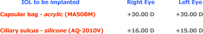
Select the appropriate polypseudophakia lens pair.
It is recommended that different IOL materials, in different locations,
be used for the creation of a polypseudophakia lens pair.
For placement in the capsular bag, an IOL with a negative shape factor
such as the Alcon MA50BM is an excellent choice because at +30.00 D, all
but 1/5th of the lens power is located on the posterior surface. For the
ciliary sulcus lens, a large diameter, low profile, round edge, biconvex
newer generation silicone IOL, such as the Staar AQ-2010V (+5.00 D to +30.0
D) or the extended power range Staar AQ-5010V (-4.00 D to +4.00 D) is recommended.
This IOL combination lowers the likelihood of pigment dispersion, iris
transillumination defects, intermittent uveitis, or secondary glaucoma,
as well as the occasional
"teeter tottering" of the sulcus placed IOL over a biconvex,
or positive shape factor, capsular bag IOL. This also lessens the occurrence
the complication of interlenticular opacifications, which has all but disappeared
following the abandonment of the original bag-bag acrylic combination.
|
Above is the IOL strategy recommended for this patient. It should be understood by all prior to this type of surgery that in spite of the very best and well-reasoned technique, the final postoperative refractive result may still end up different than expected. And because extreme axial hyperopia is far outside anything that could be considered a normal range of operation for any IOL power calculation formula, deviations from expected are not uncommon. If this is the case, the silicone lens in the ciliary sulcus position can be exchanged for one of another power, leaving the higher power acrylic lens within the capsular bag.
In situations where the refractive outcome is not certain, the practice
of operating on the non-dominant eye first is one additional way to fine-tune
the refractive outcome when it comes time for surgery on the dominant eye.
Dr. Joel Shugar in Perry Florida shared this approach with us many years
ago and we have found it very helpful when doing these very challenging
IOL power calculations.
|


