|
Zeiss IOLMaster Customer Service: 1-877-486-7473 ext.
4030
IOLMaster — Evaluation of ALM Results.
Interpretation of Axial Length Measurements
As a rule, an interference signal is produced if the measuring light is
reflected by the tear film and the retinal pigmented epithelium of the
eye. This signal is utilized for axial length measurements.
 |
|
Note
Ultrasonic biometrical instruments measure the axial length as the
distance between the cornea and the inner limiting membrane,
because the sound waves are reflected at this membrane.
To ensure that the measured values obtained with the IOLMaster
are compatible with those obtained through acoustic axial length
measurement, the system automatically adjusts for the distance
difference between the inner limiting membrane and the
pigmented epithelium.
The displayed axial length values are thus
directly comparable to those obtained by immersion ultrasound.
Deviations may nevertheless occur between the displayed axial lengths
and ultrasonic readings (particularly in the applanation procedure).
At this point the importance of re-personalizing the "lens
constants" should be stressed, because the IOLMaster is based on
a new, more precise measurement technology. Refer to the specialist
literature and publications by the originators of the IOL formulas regarding
the personalization of lens constants.
Updated information is available in the Internet at:
http://www.meditec.zeiss.com/iolmaster
and/or
http://ocusoft.de/ulib/
|
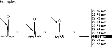
With an optimally aligned device, relatively clear eye media and weak
ametropia (< 6 D), the secondary maxima will be detected
symmetrically on each side of the actual measuring peak. These are
caused by the measuring light source used and maintain a constant
distance of approximately 0.8 mm to the measurement signal and to each
other, irrespective of the specific circumstances of the measured object.
For this reason, the secondary maxima are similarly always visible in measurements
of the supplied test eye.

Undisturbed measurement
signal
with secondary
maxima
|
|
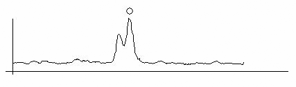
The IOLMaster measuring system is capable of resolving fine structures
on the fundus of the eye. Depending on the anatomical conditions of the measured
eye, the measuring beam may also produce interferences when reflected at
the inner limiting membrane and/or the choroid.
Indications of this are:
-
Broader (smeared) signal peaks of the measuring curve
-
Variations of approximately 150 to 350 μm in axial length data in one
measurement series and
-
Display of "Evaluation" in place of the mean value (composite
reading).
|
Such measuring curves or measurement series require immediate
verification, either between individual measurements (in ALM mode)
or in post-run editing (without the patient
in front of the IOLMaster). Interpretation or post-run editing should always
be performed with the help of the zoom function (see Zooming
Display).
|
|
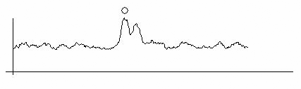
Note
The resolution of fine retinal structures is clearly distinguishable
from the previously mentioned secondary maxima, which are
further away from the multiple peaks and symmetrical to them.
The distance between the maximum peak and internal limiting
membrane or choroid is 350 μm (whereas the secondary maxima
are about 800 μm from the maximum peak!).
|
Signals From Inner Limiting Membrane (ILM)
The measuring beam is relatively often reflected at the inner limiting
membrane, likewise producing an interference signal. The respective
signal peak lies to the left of the actual measurement peak (to the
shorter axial lengths). The distance of the peak generated by the
reflection on the inner limiting membrane from the measurement peak
is between 150 and 350 µm. Both peaks can be observed separately in
a zoom view of the graph.
Double peak produced at inner limiting membrane (triple zoom)
Usually, the signal amplitude of the peak from the inner limiting membrane
is smaller than that of the interference on the pigmented epithelium. In such
a case the automatic algorithm finds the correct axial length.
 |
WARNING: Never move the measuring cursor
manually to the (left) peak produced by the inner limiting membrane (see above)! |
In rare cases the amplitude of the signal from the inner limiting membrane
may be higher than that of the reflected light from the pigmented epithelium.
In this case, the automatic peak detection will recognise the signal from
the ILM.
Signal curve with higher signal from inner limiting membrane (double zoom)
In measurement series, such individual measurements stand out by deviations
in the range of approx. 150 to 350 μm towards shorter axial lengths. The
reading can be corrected by dragging the measurement cursor in the composite
signal to the lower peak (that of the pigmented epithelium). This manipulation
is only permissible in the context of the single signals of this series of
measurements!
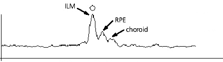
Signals From Choroid
Triple Peaks
In rare cases, the measuring beam may also be reflected by the vessels
of the choroid.
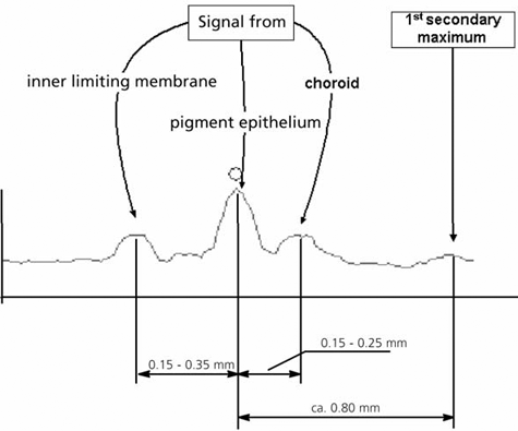
The measuring peak produced by the choroid appears shifted towards longer
axial lengths by approximately 150 to 250 μm from the peak of the pigmented
epithelium.
|
WARNING:
In the above example, the signal from the RPE (middle peak) has the highest
amplitude. The automatic peak detection system has correctly recognised
this measured value as the axial length, so that the measuring cursor may
not be moved. |
This type of rare triple peak clearly differs from the secondary maxima
produced through the light source by the distance from the RPE reflected
peak.
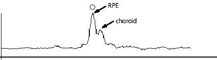
In very rare cases, depending on the anatomical conditions of the measured
eye, the signal produced by the pigmented epithelium may not be the one with
the highest amplitude.
Double peak produced by pigmented epithelium and choroid (double zoom)
The automatic peak detection system will find an axial length value that
is too short by approximately 150 to 350 μm.
|
WARNING: Following the comparison of all measured values
and curves for this eye, the measuring cursor must be moved manually to
the middle (smaller) peak produced by the RPE. This measured value is thus
corrected and shown in the display field with an asterisk. |
Double peaks
In very rare cases signals may be produced by both the pigmented epithelium
and the choroid.
Double peak produced by pigmented epithelium and choroid (double zoom)
|
WARNING:
Here again, the automatic peak detection system has placed the measuring
cursor at the correct position, as the (correct axial length) signal
from the pigmented epithelium has the greater amplitude. The
measuring cursor may not be moved. |
|
|
Note
Such a curve may only be evaluated correctly by viewing all measuring curves
of this eye. Such a curve must be clearly distinguished from double peaks
produced by the inner limiting membrane and the RPE (see Signals from
the inner limiting membrane (ILM), above).
It may be
advisable to perform further measurements. Up to 20 measurements may
be taken on one day.
Updated information is available in the Internet at:
http://www.meditec.zeiss.com/iolmaster
and/or
http://ocusoft.de/ulib/
|
|