| Zeiss IOLMaster® Online Users Instruction Manual V.5 |
|
|
|
|
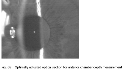
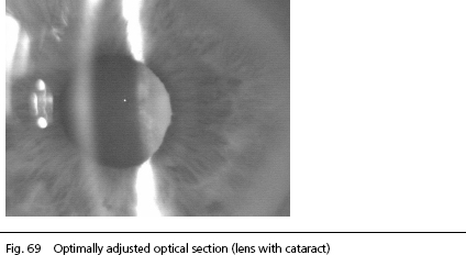
An image similar to that of a slit lamp (optical section through the anterior segment of the eye) is visible on the display. Align the IOLMaster to the patient's eye by lateral adjustment using the joystick until:
If the IOLMaster has been properly alignedthe images of the fixation point and the anterior crystalline lens will be simultaneously in focus, as they are approximately in the same plane. As a rule, the image of the fixation point lies between the image of the anterior lens and that of the cornea, if the IOLMaster is optimally aligned.
The two images above show optical sections of the right eye. The patterns to the left of the corneal image are direct reflections of the luminous light exit aperture of the lateral slit projector. These reflections are not needed for the calculation of the anterior chamber depth. They must not affect the image of the cornea (see below). At the left margin of the picture, additional reflections of the patient’s surroundings (in this case a window) are visible. Depending on the lighting conditions in the examination room, the front side of the IOLMaster as reflected by the cornea may also be visible. These artifacts do not affect the measurement of anterior chamber depth, unless the significant image details (images of cornea and crystalline lens) and the image of the fixation point are eclipsed by this extraneous light. This may be alleviated by slightly darkening the examination room.
The measurement for the anterior chamber depth on eyes with very small pupils (e.g., with glaucoma) is particularly problematic and needs some practice and experience. The anterior chamber depth of the human eye also depends on the accommodative state of the eye. This cannot be assessed from an optical section of the anterior segment.
|
|
|
|
Arizona's Top Eye Doctors - East Valley Ophthalmology provides this online information for educational and communication purposes only and it should not be construed as personal medical advice. Information published on this website is not intended to replace, supplant, or augment a consultation with an eye care professional regarding the viewer/user's own medical care. East Valley Ophthalmology's disclaims any and all liability for injury or other damages that could result from use of the information obtained from this site. Please read our full Terms, Privacy, Infringement |
Eye Doctors for Mesa, Gilbert, Chandler, AZ and surrounding areas.A few of our featured topics: |
||
Copyright
East Valley Ophthalmology. All rights reserved.
![]()