doctor-hill.com Carl Zeiss Meditec IOL Master |
IOLMaster — Anterior Chamber Depth
|
| Note Although it is not dangerous for the patient to look into the slit projector, doing so will lead to erroneous anterior chamber depth values. |
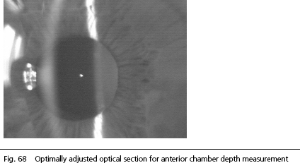
An image similar to that of a slit lamp (optical section through the anterior segment of the eye) is visible on the display. Align the IOLMaster to the patient's eye by lateral adjustment using the joystick until:
- The image of the fixation point appears optimally focused in the green square on the display.
- The image of the cornea (right eye deflected to the left, left eye to the right) is free of reflections. Not perfectly sharp is okay because of system-related lack of definition.
- The image of the anterior crystalline lens is visible in the pupil.
| Note The image of the fixation point may not lie in the image of the lens or cornea. |
If the IOLMaster has been properly alignedthe images of the fixation point and the anterior crystalline lens will be simultaneously in focus, as they are approximately in the same plane.
As a rule, the image of the fixation point lies between the image of the anterior lens and that of the cornea, if the IOLMaster is optimally aligned.
| Note The image of the fixation point should be near (but not in!) the image of the lens. |
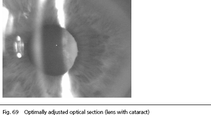
The two images above show optical sections of the right eye.
The patterns to the left of the corneal image are direct reflections of the luminous light exit aperture of the lateral slit projector. These reflections are not needed for the calculation of the anterior chamber depth. They must not affect the image of the cornea (see below).
At the left margin of the picture, additional reflections of the patient’s surroundings (in this case a window) are visible. Depending on the lighting conditions in the examination room, the front side of the IOLMaster as reflected by the cornea may also be visible. These artifacts do not affect the measurement of anterior chamber depth, unless the significant image details (images of cornea and crystalline lens) and the image of the fixation point are eclipsed by this extraneous light. This may be alleviated by slightly darkening the examination room.
 |
WARNING: The IOL Master must be adjusted very carefully for anterior chamber depth measurements. Failing to satisfy the above requirements for the measurement of the anterior chamber depth will either result in measuring errors or the measured values shown will be incorrect. Because of the complexity of the images measured, under certain circumstances measuring errors may not be recognized as such. |
The measurement for the anterior chamber depth on eyes with very small pupils (e.g., with glaucoma) is particularly problematic and needs some practice and experience.
The anterior chamber depth of the human eye also depends on the accommodative state of the eye. This cannot be assessed from an optical section of the anterior segment.
|
Note
|