Diabetic Retinopathy - Mesa, Arizona.
Diabetic Eye Disease - Early Detection Is Seriously Vital.
Diabetes affects nearly 14 million Americans. If you
have diabetes, it is important to learn as much as you can about
the effect this can have on your vision. With proper knowledge
and today's improved methods of diagnosis and treatment, you may
have a substantially reduced risk of developing serious diabetic
eye disease. We invite you to make an appointment and learn more: 480-981-6111.
|
Diabetes can cause blindness.
More than 8000 diabetic patients
in the US become blind every year. Eye disease in the form of diabetic
retinopathy is the leading cause of blindness in patients
aged 20 - 64
years. Early
detection, along with improved lifestyle, is your best protection
against permanent vision loss. By maintaining
strict control of your blood sugar and scheduling regular eye exams,
you can significantly affect the long term quality of your vision.
|
|
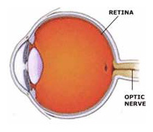
Diabetic retinopathy affects the retina and optic nerve. |
What is Diabetic Retinopathy?
If you have diabetes mellitus, your body does not regulate blood sugar
levels properly.
High blood-sugar levels can cause damage to blood vessels throughout
your body, but especially vulnerable are the blood vessels in the nerve
layer at the back of the eye, called the retina, which senses light and
helps to send images to the brain. Damage caused
by diabetes to the retinal vessels is referred
to as diabetic retinopathy.
Types of Diabetic Retinopathy.
Diabetic retinopathy can be broadly categorized into two types: non-proliferative (NPDR)
and proliferative (PDR). Diabetic retinopathy is a progressive
process. In general, there is a progression from non-proliferative
to the more severe proliferative diabetic retinopathy, but this
is not inevitable.
| |

Non-Proliferative
Diabetic Retinopathy |
Non-Proliferative Diabetic Retinopathy
Non-Proliferative Diabetic Retinopathy (NPDR), commonly
known as background retinopathy, is an early
stage of diabetic retinopathy. In this stage, tiny blood vessels within
the retina leak blood or fluid. The leaking fluid causes the retina
to swell or to form hard deposits called "exudates". Many
people with diabetes have mild NPDR, which usually does not affect
their vision. When vision is affected, it is the result of macular
edema and/or macular ischemia.
Diabetic Macular Edema
One of the earliest effects of diabetes is fluid leaking from retinal
blood vessels. This leaking causes a swelling or thickening of the macula,
a small area in the center of the retina that allows us to see fine details
clearly. The swelling is the most common cause of visual loss in diabetes.
Vision loss may be mild to severe, but even in worst cases, peripheral
vision continues to function. Any diagnosis of macular edema is an indication
that breakdown of the retinal blood vessels from diabetes is starting
to occur and requires careful monitoring.
Diabetic Macular Ischemia
Macular ischemia occurs when small blood vessels (capillaries) close.
Vision blurs because the macula no longer receives sufficient blood supply
to work properly.
| |

Proliferative
Diabetic Retinopathy
|
Proliferative Diabetic Retinopathy
Proliferative Diabetic Retinopathy (PDR) may cause more
severe vision loss than NPDR because it affects both central and
peripheral vision.
As
diabetic retinopathy begins to progress, the retinal blood vessels continue
to weaken and begin to narrow and close. As the blood vessels close,
they can no longer carry oxygen to the retinal tissue. Once deprived
of sufficient oxygen and nutrients to maintain normal health and functioning,
the retina responds by attempting to compensate by growing abnormal
new vessels blood vessels - a process called neovascularization. The
condition whereby retinal neovascularization exists is called proliferative
diabetic retinopathy.
While new blood vessel growth or neovascularization might seem like
a beneficial event, these new blood vessels are extremely fragile and
tend to break easy and hemorrhage. If left untreated, proliferative
diabetic retinopathy will in fact result in hemorrhage that leads to
scarring and ultimately to retinal detachment with profound vision loss.
Proliferative diabetic retinopathy is treated with either laser treatment
or laser treatment in conjunction with a surgical procedure called a
vitrectomy. The retinal surgeon will perform a vitrectomy in order to
remove the vitreous that has become filled with blood or scar tissue.
Sometimes it may be possible for patients to have proliferative diabetic
retinopathy and neovascularization and yet still have good vision. It
is critical that in order to maintain that good vision the neovascularization
be treated as quickly as possible even if it does appear to be causing
any vision loss.
Diagnosis and Diabetic Eye Exams
A comprehensive medical eye examination, which includes a dilated retinal
examination, is essential to detect changes inside your eye. The ophthalmologists
at East Valley Ophthalmology can often diagnose and treat serious retinopahty
before you are aware of any vision problems. Your ophthalmologist will
dilate your pupil and look inside fo the eye with special equipment
and lenses. Ancillary tests may be required depending on the severity
of retinopathy discovered. These include color fundus photography, fluorescein
angiography, and ultrasonography.
Unfortunately, because visual loss is often a late symptom of advanced
retinopathy, many patients remain undiagnosed and are examined only
after the optimal time for treatment has passed. Much of the complications
can be prevented if the retinopathy is detected early enough for treatment.
The optimal time for treatment is before you experience visual symptoms.
Patients with diabetes must be diligent and attentive to their eye
care throughout their lives. The American Diabetic Association (ADA)
recommends yearly eye examinations for all diabetic patients to detect
blood vessel leakage and the presence of diabetic retinopathy in its
earliest stages. While “screening photographs”, even through
a dilated pupil may be of some use, it is not a substitute for a comprehensive
diabetic eye examination by an ophthalmologist.
Diabetic patients may
also be more prone to other significant eye problems such as glaucoma
and cataracts and thus a comprehensive eye examination is a necessity.
Depending on the severity and progression of diabetic retinopathy, it
might be necessary to have an Intravenous Fluorescein Angiogram (IVF)
on a regular basis in order to observe and record any changes to the
retinal circulation. In general, most insurance carriers endorse and
pay for regular annual diabetic eye exams with Intravenous Fluorescein
Angiography as often as needed in order to help patients preserve vision.
High quality diabetic eye care is an important event for all diabetic
patients.
Treatment of Diabetic Retinopathy:
Treatments are very effective in reducing vision loss from this
disease. In fact, even people with advanced retinopathy have at least
a 90 percent chance of keeping their vision if they get treatment before
the retina is severely damaged. Treatments include:
Laser Treatments for Diabetic Retinopathy
Vitrectomy for Diabetic Retinopathy
·
Laser Treatments for Diabetic
Retinopathy
1) Drops are administered to dilate your pupils and numb your
eyes.
2) In some cases, the area behind your eye may be numbed by injection
as well to prevent any discomfort.
3) A special lens is placed onto your eye.
4) The lights in the office are dimmed. As you sit facing
the laser, you may see flashes of light and notice a painless
pinching sensation.
5) Your pupils will remain dilated for a few hours,
so you will need to wear dark wraparound sunglasses afterwards
and arrange for someone to drive you home.
Laser treatments are available for:
|
Proliferative Retinopathy
A procedure called scatter laser treatment dissolves the abnormal
blood vessels that form at the back of the eye. Rather than focusing
on a single spot, hundreds of tiny laser zaps shrink the abnormal blood
vessels from the center of the retina. Side vision is typically affected
by this treatment in order to save the remaining central sight and
may need repeating if new blood vessels appear.
|
|
Macular Edema
This laser surgery, called focal laser treatment, if performed early enough can
reduce vision loss from macular edema by half. During the surgery, a high-energy
beam of light is aimed directly onto the damaged blood vessels. It seals the
vessels and stops them from leaking. Sometimes, more than one treatment may be
needed to completely control the leaking fluid.
|
Studies on Laser Treatments for Diabetic Eye Disease
Three major clinical trials have conclusively
demonstrated the value of argon laser photocoagulation surgery in reducing
the rate of severe visual loss in patients with advanced diabetic retinopathy
as well as in earlier stages of the disease and for the treatment of macular
edema. Earlier surgery for severe proliferative retinopathy and vitreous
hemorrhage has also been recommended.
The aforementioned clinical trials have shown that individuals who are treated
appropriately are more likely to have better visual outcomes than those who
are not. Panretinal scatter photocoagulation using the Argon laser reduces
the rate of severe visual loss by over 50%. In this outpatient procedure
laser light is directed into the eye through a special contact lens held
on the eye while the patient sits at a slit lamp. Between 1000 and 2000 burns
a fraction of a millimeter in size are evenly placed across the entire peripheral
retina. This destruction of retinal tissue causes the proliferating vessels
to disappear by a poorly understood mechanism.
Laser treatment of clinically
significant macular edema also reduces the rate of subsequent visual loss
substantially. In this case the leaking microaneurysms are treated directly
with the argon laser to seal them and allow the edema to be resorbed preventing
further vision loss. Vitrectomy and retinal detachment surgery may become
necessary in cases of persistent, non-clearing vitreous hemorrhage and for
traction retinal detachment due to scarring.
|
·
Vitrectomy
Instead of laser surgery, an eye operation called a vitrectomy may be needed
to restore sight. A vitrectomy is performed in cases that have a lot of blood
in the vitreous. It involves removing the cloudy vitreous and replacing it with
a special salt solution.
|
1) Studies show that people who have a vitrectomy soon after a large hemorrhage
are more likely to protect their vision.
2) Early vitrectomy is especially effective in people with insulin-dependent
diabetes, who may be at greater risk of blindness from a hemorrhage into the
eye.
3) Vitrectomy is often done under local anesthesia, which means that you will
be awake during the operation. Tiny incisions are made in the sclera, or white
of the eye. Then, small instruments are placed into the eye that remove the vitreous
and replace it with the salt solution.
4) Your eye will be red and sensitive. You may be able to return home soon afterwards,
or you may be asked to stay in the hospital overnight. An eye patch is required
thereafter, for a few days or weeks to protect the eye, as well as medicated
eye drops to protect against infection.
Ideal Treatment for Diabetic Retinopathy
The ideal treatment fo diabetic retinopathy is
to prevent the development of the disease as much as possible, as it
is possible to maintain vision and prevent severe vision loss for many
patients. Strict control of your blood sugar will significantly reduce
the long-term risk of vision loss from diabetic retinopathy. If high
blood pressure and kidney problems are present, they need to be treated.
The eye specialists of East Valley Ophthalmology perform advanced
technology diagnostic testing and treatment, as well as taking
the time necessary to provide each patient with information needed
to fully understand their condition and to achieve their best possible
visual outcome.
If you would like further information, please call our office at:
480-981-6111
East Valley Ophthalmology
Eye Doctors - Mesa, ArizonaIf you or a family member
or friend have not had a recent routine eye examination, have a specific eye condition that needs addressing, or are looking for
an eye specialist or professional eye consultant please take a moment to Request an Appointment.
|
