
A-scan biometry by applanation requires that the ultrasound probe be placed directly on the corneal surface. This can either be done at the slit lamp or by holding the ultrasound probe by hand.
Even in the most experienced hands, some compression of the cornea is unavoidable; this typically ranges from 0.14 mm to 0.28 mm.
The popularity of the applanation method is due to its apparent speed.
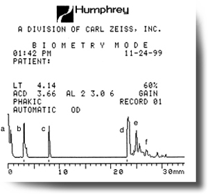 Figure A – Phakic axial length measurement using the applanation method.
- Initial spike (probe tip and cornea)
- Anterior lens capsule
- Posterior lens capsule
- Retina
- Sclera
- Orbital fat
Note: When echoes b, c, and d are high and steeply rising, the ultrasound beam is most likely on axis. The scleral echo should easily be identified, and the orbital fat echoes should descend quickly and at a steep angle. If there are no scleral or orbital fat echoes visible, the ultrasound beam is most likely aligned with the optic nerve rather than the macula.
 Figure B – Note the typical variations in applanation measurements.
Measurements taken by the applanation method will often show variability from one to the next, resulting from inconsistent corneal compression, and this variability can be observed even under the most experienced guidance.
To avoid this, switch to the immersion technique, as described below.
How to learn more about a-scan techniques:
We highly recommend the book A-scan Axial Length Measurements by Sandra Frazier Byrne.
There is an excellent, national certification program in Ophthalmic Biometry available for your technicians:
